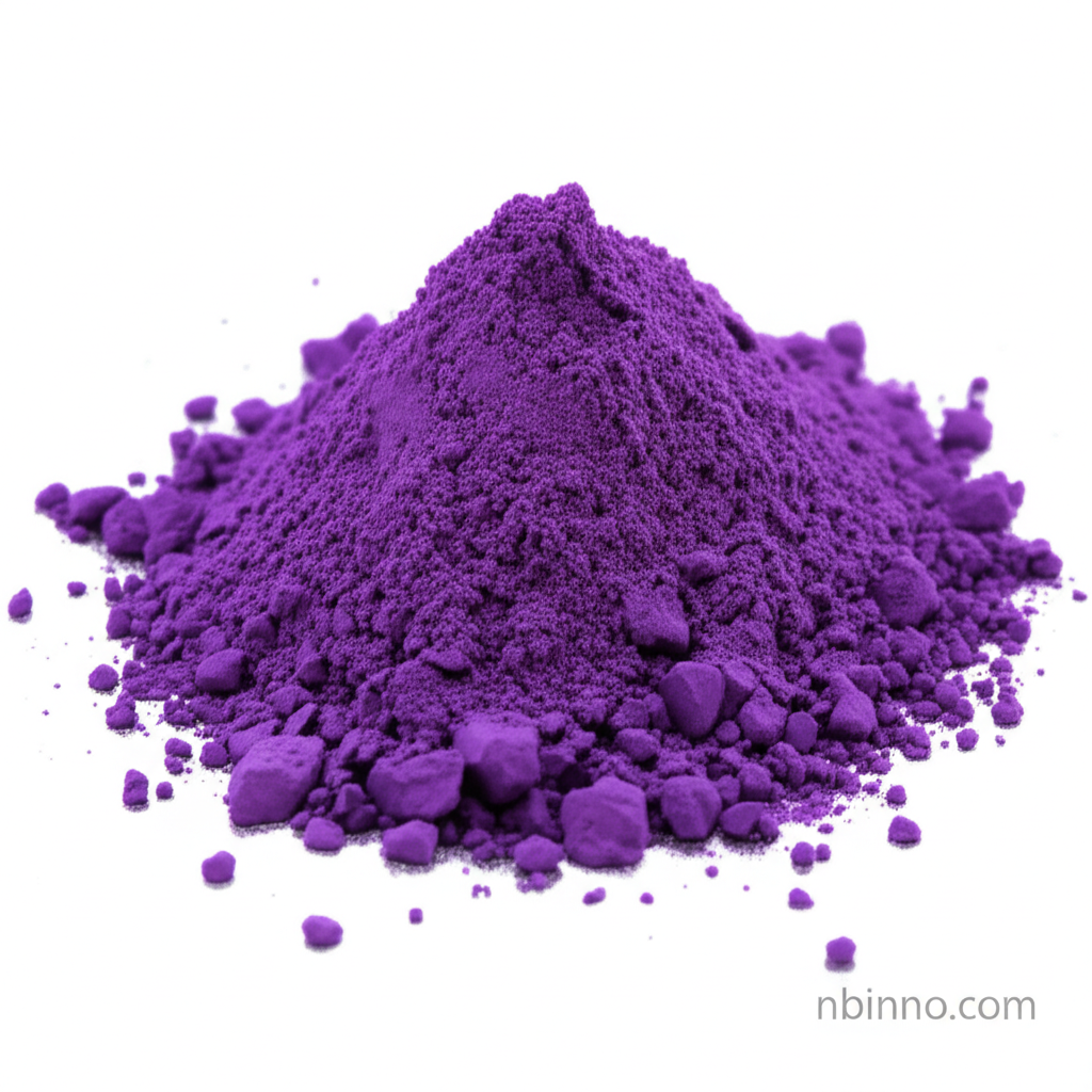
Light Green SF Yellowish: Essential for Precise Histological Staining
Discover the versatility of Light Green SF Yellowish, a key dye for detailed microscopic analysis and tissue visualization.
Get a Quote & SampleProduct Core Value

Light Green SF Yellowish
Light Green SF Yellowish is a crucial biological stain, highly valued for its role as a counterstain in various histological and cytological applications. Its ability to differentiate cellular components and tissues makes it indispensable for accurate microscopic examination.
- Explore the use of Light Green SF Yellowish as a vital counterstain in cytology for enhanced visualization.
- Understand how Acid Green 5 serves as a reliable substitute for aniline blue in Masson's trichrome staining procedures.
- Learn about the application of CAS 5141-20-8 in accurately staining collagen fibers for detailed connective tissue analysis.
- Discover the importance of Color Index 42095 in diagnostic staining, particularly within Papanicolaou (PAP) stains for cellular differentiation.
Key Advantages Offered
Enhanced Cellular Differentiation
Leverage the precise staining capabilities of Light Green SF Yellowish, a critical component for differentiating cytoplasmic and extracellular structures, aiding in the identification of various cell types.
Versatile Application Spectrum
Benefit from the stain's adaptability, serving effectively as a counterstain in cytology and as a direct substitute for aniline blue in established histological protocols like Gomori's step.
Reliable Quality and Consistency
Ensure reproducible results with a dye that meets stringent quality standards, suitable for both routine laboratory use and specialized research, contributing to accurate histological staining.
Key Applications
Cytology Counterstaining
As a key counterstain, it provides essential contrast in cytological preparations, making cellular details more discernible for diagnostic purposes.
Histological Tissue Staining
It's widely used in histology to stain cytoplasmic and extracellular components, offering a distinct green hue that aids in the comprehensive analysis of tissue morphology.
Collagen Fiber Visualization
This dye effectively stains collagen fibers, serving as a valuable alternative to aniline blue in trichrome staining techniques, crucial for studying connective tissues.
Diagnostic Staining Protocols
Integral to methods like the Papanicolaou (PAP) stain, it enhances the visualization of cellular structures, contributing to accurate diagnoses in medical laboratories.
